
Hair Scan in Islamabad
Hair Scan is a non-invasive digital assessment of the scalp and hair that uses magnification, dermoscopy or video-trichoscopy to examine hair shafts, follicular openings, hair-density patterns and visible scalp changes in greater detail than is possible with the naked eye.
A Hair Scan can help support assessment of concerns such as pattern hair loss, excessive shedding, patchy hair loss, scalp scaling and selected hair-shaft abnormalities. It is an assessment tool rather than a treatment, and scan findings should be interpreted together with medical history and clinical examination. Blood tests, fungal testing or scalp biopsy may still be required when clinically indicated. Trichoscopy is recognised as a useful non-invasive technique for evaluating hair and scalp disorders.
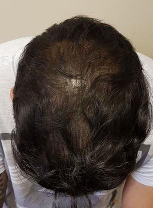
What is Hair Scan?
Hair Scan is a patient-friendly term commonly used for digital examination of the scalp and hair using magnification.
In clinical dermatology, a more specific term is trichoscopy.
Trichoscopy involves examination of the scalp and hair using a:
- Handheld dermatoscope
- Digital dermatoscope
- Video-trichoscope
- High-magnification scalp camera
Depending on the system, images can be enlarged and displayed on a monitor so that the clinician can assess structures that may be difficult to see during routine examination.
Trichoscopy can visualise features including:
- Hair-shaft thickness
- Variation in hair diameter
- Vellus or miniaturised hairs
- Broken hairs
- Follicular openings
- Empty follicles
- Yellow dots
- Black dots
- White dots
- Scaling
- Perifollicular changes
- Scalp pigmentation
- Selected vascular patterns
DermNet describes trichoscopy as a valuable non-invasive technique that can help evaluate most hair and scalp disorders and allows digital images to be stored for comparison over time.
What Is Hair Scan Used For?
Hair Scan may be used as part of the assessment of:
- Male pattern hair loss
- Female pattern hair loss
- Excessive hair shedding
- Alopecia areata
- Telogen effluvium
- Patchy hair loss
- Traction alopecia
- Selected scarring alopecias
- Hair breakage
- Hair-shaft disorders
- Dandruff and scalp scaling
- Selected inflammatory scalp conditions
- Treatment progress
The scan can help a clinician recognise patterns that suggest different types of hair loss.
For example, trichoscopy can assist in distinguishing scarring from non-scarring alopecia and can help differentiate conditions such as androgenetic alopecia and telogen effluvium.
A Personalized Approach to Hair Scan
Hair loss and scalp concerns can have many different causes, so a Hair Scan should be used as part of an individual clinical assessment rather than treated as an automatic diagnosis or treatment recommendation. Digital magnification can provide valuable information about hair density, follicular openings, hair-shaft diameter, miniaturisation and scalp changes, but these findings need to be interpreted within the patient's overall clinical picture.
Before performing or interpreting a Hair Scan, Dr. Samavia reviews each patient individually, considering when the hair loss started, whether it is gradual or sudden, the pattern of thinning or shedding, family history, scalp symptoms, current medications, medical history, previous hair treatments and any relevant nutritional, hormonal or systemic factors.
The scalp and hair are then examined using appropriate magnification or trichoscopic imaging. Depending on the concern, Dr. Samavia may evaluate hair-density patterns, variation in hair-shaft diameter, follicular miniaturisation, broken hairs, follicular openings, scaling, redness, perifollicular changes and other clinically relevant findings.
A Hair Scan can help support the assessment of conditions such as androgenetic alopecia, telogen effluvium, alopecia areata, traction alopecia, selected scarring alopecias and inflammatory or scaling scalp disorders. However, the scan does not independently identify every cause of hair loss.
If the clinical history or scan findings suggest another contributing problem, the personalised assessment plan may also include blood investigations, iron or thyroid testing, fungal testing, hair-pull assessment, dermatological evaluation or scalp biopsy when clinically indicated.
The purpose of the assessment is to determine what is actually happening before selecting treatment. A Hair Scan should therefore help guide whether the patient may benefit from medical hair-loss treatment, PRP or PRF, low-level laser therapy, scalp treatment, hair transplantation, observation or another clinically appropriate approach, rather than automatically leading to a particular procedure.
Monitoring Hair and Scalp Changes
One of the important advantages of digital Hair Scan or trichoscopy is the ability to create a baseline record that can be compared with future assessments.
Where appropriate, Dr. Samavia may document selected scalp areas using standardised photographs or magnified images and record findings such as hair density, hair-shaft variation, follicular miniaturisation and scalp condition.
Repeat Hair Scans may then be used during follow-up to help assess whether:
-
Hair density appears stable or is changing
-
Follicular miniaturisation is progressing
-
Existing hairs appear thicker
-
Additional thinning is occurring
-
Scalp inflammation or scaling has improved
-
A treatment plan appears to be producing a meaningful clinical response
Because hair grows slowly, repeat assessments need to be performed at clinically appropriate intervals. Comparing scans too frequently may create misleading impressions because meaningful changes in hair growth generally develop over months rather than days or weeks.
Dr. Samavia interprets follow-up findings together with the patient's symptoms, clinical examination and treatment history. Digital measurements and software-generated results are supportive tools and should not be treated as infallible diagnoses or guarantees of future hair growth.
If you are experiencing hair thinning, excessive shedding, patchy hair loss or persistent scalp concerns and are considering a Hair Scan in Islamabad, you can book a consultation with Dr. Samavia for an individual hair and scalp assessment and a personalised diagnostic and treatment plan.
What concerns can it address?
Hair Density
Digital examination may help document differences in visible hair density between scalp areas.
For pattern hair loss, comparison between areas such as the frontal and occipital scalp can be particularly useful.
Hair-Shaft Diameter
Healthy scalp areas generally contain terminal hairs of relatively consistent diameter.
In androgenetic alopecia, progressive follicular miniaturisation can result in increasing variation between thick terminal hairs and finer hairs.
A 2024 systematic review found hair-diameter variability to be one of the most common trichoscopic findings in androgenetic alopecia.
Hair Miniaturisation
Androgenetic alopecia gradually converts some terminal hairs into shorter, finer, miniaturised hairs.
A Hair Scan can help identify this pattern.
Other findings associated with androgenetic alopecia can include:
- Increased vellus hairs
- Reduced multi-hair follicular units
- Peripilar signs
- Hair-diameter diversity
These findings must be interpreted within the overall clinical picture.
Follicular Openings
Trichoscopy allows examination of follicular openings.
This can be important because loss of follicular openings may raise concern for certain types of scarring alopecia, where early diagnosis can be particularly important.
Broken Hairs
The scan may show:
- Broken hairs
- Black dots
- Tapered hairs
- Abnormal hair-shaft patterns
These findings may provide clues in conditions such as:
- Alopecia areata
- Trichotillomania
- Tinea capitis
- Hair-shaft disorders
Trichoscopy is commonly used to help differentiate causes of localised non-scarring hair loss.
Scalp Scaling and Inflammation
Magnification may reveal:
- Scaling
- Follicular scaling
- Redness
- Perifollicular changes
- Crusting
- Other scalp abnormalities
These findings may support assessment of inflammatory or infectious scalp conditions.
Potential benefits
Potential benefits of a Hair Scan include:
- Non-invasive assessment
- No injections
- No anaesthesia
- No radiation
- No downtime
- Magnified assessment of hair and scalp
- Can document hair miniaturisation
- May help distinguish different hair-loss patterns
- Can support early recognition of selected alopecias
- Images can be stored for future comparison
- Can help monitor treatment response
- Can help determine whether further investigation is required
- May help identify an appropriate scalp-biopsy site when biopsy is necessary
Published reviews describe trichoscopy as useful not only for diagnosis but also for treatment monitoring and, in selected cases, choosing the most useful area for scalp biopsy.
Limitations and realistic expectations
Hair Scan is an assessment method, not a complete diagnosis by itself.
It cannot automatically determine every cause of hair loss.
A scan cannot independently tell a patient with certainty:
- Why iron-related hair shedding is occurring
- Whether thyroid disease is present
- Whether hormone abnormalities exist
- Whether nutritional deficiency is present
- Whether a medication is causing hair loss
- Whether every scalp condition is fungal
- Whether hair will definitely regrow
- Which treatment will guarantee success
Medical history remains essential.
Depending on the suspected cause, additional investigations may include:
- Blood tests
- Thyroid testing
- Iron studies
- Other laboratory investigations
- Fungal testing
- Hair-pull testing
- Scalp biopsy
Research reviewing hair-loss assessment methods emphasises that no single evaluation method is ideal for every patient. Different techniques provide different information and must be interpreted appropriately.
Patients should therefore be cautious of claims such as:
- “Hair Scan diagnoses every type of hair loss”
- “AI scan gives 100% accurate diagnosis”
- “Hair Scan predicts exactly how many hairs you will regrow”
- “One scan tells us the perfect treatment”
- “No doctor assessment is required”
- “Hair Scan replaces blood tests”
These claims are not realistic.
Hair Scan for Male Pattern Hair Loss
Male pattern hair loss, or androgenetic alopecia, usually develops gradually.
Typical changes can include:
- Frontal recession
- Temporal recession
- Crown thinning
- Progressive miniaturisation
In many men, history and clinical examination may be sufficient to recognise classic androgenetic alopecia.
Trichoscopy can provide additional information by demonstrating features such as:
- Hair-diameter variability
- Miniaturised hairs
- Vellus hairs
- Changes in follicular units
- Peripilar signs
A 2024 systematic review found hair-diameter variability in approximately 94% of assessed androgenetic alopecia cases across the included literature, making it an important trichoscopic feature.
Hair Scan for Female Pattern Hair Loss
Female pattern hair loss can sometimes be more diagnostically challenging.
Women may develop:
- Wider central part
- Reduced crown density
- Diffuse frontal or central thinning
- Increased visibility of scalp
Several other causes of diffuse shedding can produce a similar appearance.
The 2024 systematic review of androgenetic alopecia trichoscopy noted that while clinical assessment may often establish the diagnosis in men, evaluation in women can particularly benefit from trichoscopic assessment.
A clinician may also consider whether additional investigation is required for:
- Iron deficiency
- Thyroid disease
- Hormonal abnormalities
- Recent illness
- Medication changes
- Nutritional factors
- Telogen effluvium
depending on the history.
Hair Scan for Alopecia Areata
Alopecia areata commonly causes sudden patches of non-scarring hair loss.
Trichoscopy may reveal characteristic features including selected combinations of:
- Yellow dots
- Black dots
- Broken hairs
- Tapered hairs
- Short regrowing hairs
These findings can help support the diagnosis and assess disease activity.
However, clinical interpretation is still required.
Hair Scan for Telogen Effluvium
Telogen effluvium typically causes increased diffuse shedding.
Possible triggers include:
- Significant illness
- Fever
- Surgery
- Major physiological stress
- Rapid weight change
- Nutritional deficiency
- Certain medicines
- Postpartum hormonal changes
Trichoscopy may help distinguish diffuse shedding from patterned miniaturisation, although history and appropriate laboratory assessment can be particularly important.
Hair Scan does not identify the systemic trigger by itself.
Hair Scan for Scalp Conditions
Hair Scan can also magnify the scalp surface.
This may allow clearer assessment of:
- Dandruff
- Scaling
- Follicular redness
- Crusting
- Broken hairs
- Selected inflammatory patterns
Possible scalp conditions can include:
- Seborrheic dermatitis
- Psoriasis
- Tinea capitis
- Inflammatory alopecias
- Other scalp disorders
Digital magnification may help identify patterns, but a scan alone should not be used to self-diagnose or automatically prescribe treatment.
Who may be suitable?
Hair Scan may be useful for adults or appropriate younger patients with:
- Gradual hair thinning
- Increased shedding
- Receding hairline
- Crown thinning
- Widening hair part
- Patchy hair loss
- Unexplained hair breakage
- Scalp scaling
- Persistent scalp symptoms
- Previous hair-loss treatment
- Interest in documenting baseline hair density
It may also be useful before treatments such as:
- PRP
- PRF
- Medical hair-loss therapy
- Low-level laser treatment
- Hair transplantation
when the purpose is to document baseline findings.
However, performing a scan does not automatically mean that one of these treatments is appropriate.
Who may require additional assessment?
Additional medical assessment is particularly important when there is:
- Sudden severe shedding
- Rapidly progressive hair loss
- Patchy hair loss
- Painful scalp
- Significant scalp inflammation
- Pustules
- Scarring
- Loss of follicular openings
- Significant itching
- Hair loss with systemic symptoms
- Hair loss after starting medication
- Hair loss in children
- Suspected fungal infection
Early recognition of scarring alopecia is important because permanent follicular destruction can occur in some forms.
Trichoscopy may help distinguish scarring and non-scarring hair-loss patterns, but further evaluation or biopsy may sometimes be necessary.
How to prepare
Hair Scan generally requires very little preparation.
Your clinic may ask you to:
- Avoid applying hair fibres immediately beforehand.
- Avoid heavy scalp oils on the day of assessment.
- Avoid excessive styling products.
- Bring information about current hair products.
- Bring a list of medications and supplements.
- Mention recent hair treatments.
- Mention previous PRP, PRF or hair-transplant procedures.
- Mention family history of hair loss.
- Describe when shedding began.
Do not stop normal hair-loss medication simply for a Hair Scan unless specifically instructed.
Tell your clinician about:
- Minoxidil use
- Finasteride or dutasteride where relevant
- Hormonal medication
- Supplements
- Previous transplant
- PRP or PRF
- Recent illness
- Weight loss
- Pregnancy or childbirth where relevant
- Nutritional problems
- Thyroid disease
- Previous blood-test abnormalities
because these details may help interpretation.
How the Hair Scan works
1. Medical and hair-loss history
The practitioner asks about:
- When hair loss started
- Whether it is gradual or sudden
- Shedding
- Family history
- Medication
- Medical conditions
- Previous treatment
2. Visual scalp examination
The overall pattern of thinning is assessed.
This can include:
- Frontal hairline
- Temples
- Mid-scalp
- Crown
- Sides
- Occipital donor area
3. Magnified imaging
A dermatoscope, trichoscope or digital scalp camera is positioned against or close to selected scalp areas.
Magnification can reveal details of:
- Hair shafts
- Follicular units
- Scalp skin
- Scaling
- Pigmentation
- Miniaturisation
Traditional trichoscopy commonly uses magnification in approximately the 20× to 160× range, although modern digital systems may offer additional magnification capabilities.
4. Comparison between areas
The clinician may compare affected scalp with a reference area.
For pattern hair loss, frontal or central scalp findings may be compared with the occipital scalp.
5. Digital images
Selected images may be saved.
This allows future comparison after treatment.
6. Clinical interpretation
Findings are interpreted together with examination and history.
7. Additional testing if required
The clinician may recommend further investigations instead of immediately prescribing an aesthetic treatment.
What can digital Hair Scan software measure?
The capabilities vary by device.
Depending on the system, software may estimate:
- Hair density
- Hair count within an imaged area
- Hair-shaft diameter
- Variation in shaft diameter
- Terminal-to-vellus hair characteristics
- Number of hairs per follicular unit
- Scalp coverage
- Selected scalp-surface characteristics
More advanced photographic techniques such as phototrichograms or systems such as TrichoScan can quantify selected hair-growth characteristics.
However, software output should not be treated as an infallible medical diagnosis.
Hair-count estimates can be influenced by:
- Image quality
- Hair length
- Hair colour
- Lighting
- Scalp contrast
- Device calibration
- Area selected
- Operator technique
- Software algorithm
Consistent photography and examination technique are especially important when scans are being compared over time.
Results and treatment timeline
A Hair Scan provides information immediately.
Initial assessment
The first scan can establish a visual baseline.
This may document:
- Hair density
- Miniaturisation
- Hair-shaft variation
- Scalp condition
- Pattern of thinning
Follow-up assessment
Repeat imaging may be useful after a sufficient treatment interval.
Comparing scans too frequently can be misleading because clinically meaningful hair-growth changes take time.
For many hair-loss treatments, meaningful assessment is performed over months rather than days or weeks.
Treatment monitoring
Repeat scans can potentially help show whether:
- Hair density appears stable
- Miniaturisation is progressing
- Some hairs appear thicker
- Additional thinning is occurring
- The scalp condition has changed
Digital image storage is one recognised advantage of video-trichoscopy because serial images can be used for follow-up.
Risks and side effects
Hair Scan itself is generally very low risk because it is non-invasive.
There are:
- No injections
- No surgical wounds
- No laser energy
- No radiation
- No conventional recovery period
Possible minor issues can include:
- Temporary pressure from contact devices
- Mild disturbance of styled hair
- Rare irritation from interface gel or cleaning products
The greater risk is usually misinterpretation, rather than physical injury.
For example, poor interpretation could lead to:
- Incorrect diagnosis
- Unnecessary treatment
- Missing scarring alopecia
- Delaying treatment for fungal disease
- Assuming nutritional hair loss is genetic
- Starting cosmetic procedures before identifying an underlying medical cause
The scan should therefore be interpreted in clinical context.
Recovery and aftercare
After the Hair Scan
No recovery is normally required.
Patients can generally immediately:
- Wash their hair
- Style their hair
- Return to work
- Exercise
- Continue normal activities
If an interface gel or solution has been used, it can be removed after the assessment.
The more important next step is following the assessment plan.
Depending on findings, this may involve:
- Observation
- Medical skincare for the scalp
- Blood investigations
- Hair-loss medication
- Dermatology assessment
- PRP or PRF consideration
- Low-level laser therapy
- Hair transplantation assessment
- Another targeted treatment
A Hair Scan should help determine whether treatment is appropriate rather than automatically being used to sell a procedure.
Alternatives and comparisons
Hair Scan vs Hair-Loss Blood Tests
These tests provide different information.
Hair Scan / Trichoscopy examines the hair and scalp directly.
It may reveal:
- Miniaturisation
- Hair-shaft changes
- Follicular patterns
- Scalp changes
Blood tests assess internal factors that may contribute to hair loss.
Depending on history, these may evaluate factors such as:
- Iron status
- Thyroid function
- Selected nutritional abnormalities
- Hormonal concerns
One cannot automatically replace the other.
Hair Scan vs Trichoscopy
In many clinics, Hair Scan is a commercial or patient-friendly name.
Trichoscopy is the established medical term for dermoscopic examination of the hair and scalp.
A high-quality Hair Scan should therefore ideally involve more than taking a normal photograph.
It should use appropriate magnification and interpretation of clinically meaningful scalp and hair structures.
Hair Scan vs TrichoScan
These terms are not identical.
Trichoscopy refers broadly to magnified dermoscopic examination.
TrichoScan is a computer-assisted phototrichogram method used to quantify selected parameters of hair growth and density.
Different clinics may use different equipment.
Patients should therefore ask what technology is actually included when a service is advertised as a “Hair Scan.”
Hair Scan vs Hair Transplant Assessment
A Hair Scan can be useful during assessment for hair transplantation, but transplant planning requires substantially more information.
A proper transplant assessment considers:
- Diagnosis
- Donor density
- Donor miniaturisation
- Hair calibre
- Hair-loss pattern
- Future progression
- Age
- Existing medical treatment
- Scalp condition
- Realistic graft requirements
A digital scan alone cannot determine whether a patient is a good transplant candidate.
What affects the cost?
The cost of Hair Scan may depend on:
- Type of imaging device
- Trichoscopy versus basic scalp camera
- Number of scalp areas examined
- Digital density analysis
- Medical consultation
- Written assessment
- Comparison with previous scans
- Follow-up monitoring
- Additional hair-loss investigations
A simple salon scalp-camera examination should not automatically be considered equivalent to clinician-interpreted trichoscopy.
When comparing services, patients may wish to ask whether the assessment includes:
- Clinical hair-loss history
- Medical scalp examination
- Magnified trichoscopy
- Digital image storage
- Comparison between scalp regions
- Diagnosis or differential diagnosis
- Recommendations for further testing when required
The value of Hair Scan comes primarily from appropriate interpretation, not simply from the magnification level of the camera.
Patient experiences
Reviews describe individual experiences and do not predict another patient's result.
I had a wonderful experience consulting with Dr. Samaviya Ejaz. She is an incredibly impressive and professional dermatologist. Her clinic is beautiful and has a calming atmosphere.
Umar Shah
Read on GoogleI had my hair fall consultation with Dr Samavia. She thoroughly explained the treatment. After PRF sessions my hair fall stopped and density improved. Highly recommend.
Moneeb Mumtaz
Read on GoogleI have had a few treatments at this clinic and I am happy with the outcomes. Dr. Samavia and her team are kind, professional, and trustworthy.
Sheikh Yousuf Kareem
Read on GoogleVisit Dr Samavia at Facetime Clinic
Use the verified Google profile for clinic directions, business details and linked patient reviews.
Clinical references
- Trichoscopy: A Complete OverviewDermNet · accessed 30 August 2026
- Trichoscopy of Androgenetic Alopecia: A Systematic ReviewJournal of Clinical Medicine / PubMed · accessed 30 August 2026
- Trichoscopy: An UpdateActas Dermo-Sifiliográficas / PubMed · accessed 30 August 2026
- Trichoscopy: A New Method for Diagnosing Hair LossJournal of Drugs in Dermatology / PubMed · accessed 30 August 2026
- Evaluation and Diagnosis of the Hair Loss Patient: Trichoscopic and Laboratory EvaluationsJournal of the American Academy of Dermatology / PubMed · accessed 30 August 2026
- Hair Evaluation Methods: Merits and DemeritsInternational Journal of Trichology / PubMed · accessed 30 August 2026
Hair Scan FAQs
What does a Hair Scan show?
A Hair Scan can magnify the hair and scalp to show features such as hair-shaft thickness, variation in hair diameter, miniaturised hairs, follicular openings, scaling, broken hairs and selected scalp changes. These findings can help assess different forms of hair loss, but they must be interpreted together with clinical history and examination.
Can a Hair Scan diagnose hair loss?
A Hair Scan can strongly support diagnosis of many hair and scalp disorders, particularly when proper trichoscopy is used. However, it cannot independently identify every cause of hair loss. Some patients still require blood tests, fungal testing or scalp biopsy depending on their symptoms and examination.
Can a Hair Scan detect male or female pattern hair loss?
Yes. Trichoscopy can identify characteristic findings of androgenetic alopecia such as variation in hair-shaft diameter, miniaturised or vellus hairs and changes in follicular units. These findings can be particularly useful when the diagnosis is early or when diffuse female hair loss has several possible causes.
Is Hair Scan painful?
No. Hair Scan is normally non-invasive and painless. A handheld or digital magnifying device is placed against or close to selected parts of the scalp. There are no injections, anaesthesia or conventional downtime.
Can Hair Scan tell which hair treatment will work for me?
It can help identify the type and pattern of hair loss, which is important when choosing treatment, but it cannot guarantee that a specific therapy will work. Treatment decisions may also depend on age, medical history, blood tests, severity, duration of hair loss and whether follicles are still capable of producing hair.
Other Hair Procedures guides
Book a consultation in Islamabad to review suitability, alternatives, expected recovery and individual risks.